Diastasis recti abdominis (DRA) is the widening of the midline that separates the two rectus abdominis muscles. Between the two muscles runs a thick white fascia, the linea alba, that holds them together and has a normal width of about 2.7 cm [1]. During pregnancy this line is susceptible to horizontal stretch mainly due to hormonoelastic changes of the connective tissues and the mechanical loads that stress this particular area as the fetus grows.
The DRA usually happens at the second semester of pregnancy. At the third semester 66% to 100% of pregnant women have DRA [2, 3, 4] and 53% have DRA immediately after childbirth [5].
There are three ways to measure DRA. A) Finger width, B) Imaging ultrasound and C) calipers. Studies have shown that all three methods are reliable. The most reliable method is ultrasound imaging but in clinical settings the finger width and calipers are more practical [6,7].
Risk factors for DRA are age (older than 34 years old), multiple pregnancy (e.g. twins), big fetuses and the C-section [8].
Clinical Course of DRA
Most of the reduction of DRA usually takes place in the 8 first weeks postpartum [9] and it’s prevalence more than 1 year postpartum has been shown to reach a 32,6% of the women that were diagnosed with DRA as they were pregnant [10].
DRA has been correlated with lumbar and pelvic pain [11], reduction of strength and stamina of the abdominal muscles even in women that followed aerobic exercise programs during pregnancy [12] and with urogynecological dysfunction [13]. The latter is very interesting because we see that women that had DRA due to pregnancy (-ies) where found to have pelvic floor issues and urinal incontinence when they reached menopause.
There seem to be two types of women that develop DRA due to pregnancy. The first type are these women that, through exercise, managed to develop ways to overcome the functional limitations that appear with DRA and are capable of transferring mechanical loads through their trunk successfully, with or without reducing their DRA. The second type are women that have DRA that perseveres and have not managed to develop coping strategies, leading to lumbar pain and movement dysfunction mainly regarding their trunk [14]. The women that fit to the second type have to do regular and specific exercise and if the dysfunction and pain do not get better then surgery is the next option [15, 16].
DRA and Therapeutic Exercise
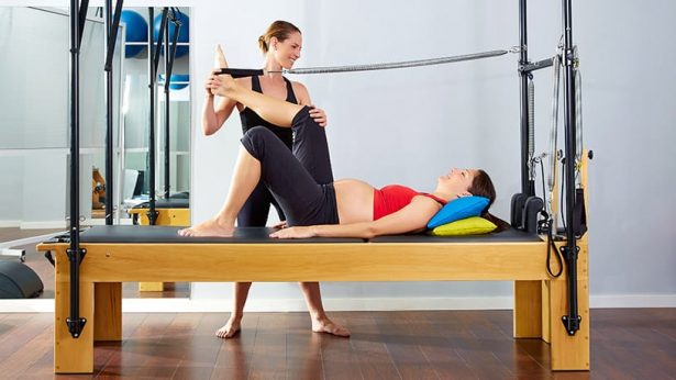
Strengthening of the abdominal muscles can help women with DRA reduce the width of the linea alba and overcome their functional limitations and pain. Another benefit is the aesthetic result of exercise that will boost up the women’s confidence and moral by reducing the gap between their abdominals. The exercises have to target the rectus abdominus muscle and the other muscles of the trunk. The sessions are more effective if they are supervised by experts such as clinical Pilates instructors.
Exercise during pregnancy
Compared to women that do not perform any kind of exercise during pregnancy, those that exercise during their pregnancy have 33,3% chance of not developing DRA [17].
However most impressive are the results from exercise in the cases of women that have already developed DRA during pregnancy. There are statistics that show that women that followed systematically exercise regimes during pregnancy developed the smallest DRA and for these women DRA correction time was the least [18, 8]. Even regular walking during pregnancy has been shown to reduce chances of developing DRA [5].
Exercise after pregnancy
Supervised therapeutic exercise such as Pilates impressively can reduce the size of the DRA of women after childbirth. Clinical Pilates focuses a lot on the correct way of contracting the abdominal wall and the pelvic floor for women with DRA, leading to better functional and aesthetic results. In addition to exercise special taping tacniques can enhance the therapeutic results [19, 20, 21, 22].
Conclusions
In conclusion we see that DRA is a very common issue that has discomforting effects on women after pregnancy, such as limitations in the function of the musculoskeletal system that has to do with distribution of mechanical load when walking, running, bending etc, but also the pelvic floor muscles and of curse lumbar pain. We know that therapeutic exercise during pregnancy can reduce the chances of one acquiring DRA but also can reduce the DRA itself with remarkable results in function and appearance of the abdomen. It is therefore strongly advised that pregnant women after consulting their obstetrics specialist should follow a supervised exercise program that focuses on strength and control of the trunk and the pelvic floor. The most suitable type of exercise for targeting the aforementioned goals is Clinical Pilates.
References
- Rath AM, Attali P, Dumas JL, Goldlust D, Zhang J, Chevrel JP. Theabdominal linea alba: an anatomo-radiologic and biomechanical study.Surg Radiol Anat 1996;18:281–8.
- Boissonnault JS, Blaschak MJ. Incidence of diastasis recti abdominisduring the childbearing year. Phys Ther 1988;68:1082–6.
- Hanneford R, Tozer J. An investigation of the incidence, degree andpossible predisposing factors of the rectus diastasis in the immedi-ate post-partum period. J Nat Obstet Gynaecol Special Group AustPhysiother Assoc 1985;4:29–32.
- P, Pascoal A, Carita A, Bo A. Prevalence and risk factors of diastasis recti abdominis for late pregnancy to 6 months postpartum, and relationship with lumbo-pelvic pain. Manual Therapy 2015; 20: 200-205.
- Candido G, Lo T, Janssen P. Risk factors for diastasis of therecti abdominis. J Assoc Chart Physiother Womens Health 2005;97:49–54.
- Mota P, Pascoal AG, Sancho F, Carita AI, Bo K. Reliability of theinter-rectus distance easured by palpation. Comparison of palpationand ultrasound measurements. Man Ther 2013;18:294–8.
- Turan V, Colluoglu C, Turkyilmaz E, et al. Prevalence of diastasis recti abdominis in the population of young multiparous adults in Turkey. Ginekol Pol 2011;82:817–21.
- Lo T, Candido G, Janssen P. Diastasis of the recti abdominis inpregnancy: risk factors and treatment. Physiother Can 1999;51:32–6,44.
- Coldron Y, Stokes MJ, Newham DJ, Cook K. Postpartum characteristics of rectus abdominis on ultrasound imaging. Man Ther 2008 May;13(2):112e21.
- Sperstad J, Tennfjord M, Hilde G, Ellström M. Diastasis recti abdominis during pregnancy and12 months after childbirth: prevalence, risk factors and report of lumbopelvic pain. Br Jour Sports Med 2016; 50:1092-1096.
- Parker MA, Millar AL, Dugan SA. Diastasis rectus abdominis and lumbo-pelvic pain and dysfunction—are they related? J Womens Health Phys Ther 2009;33:15–22.
- Gilleard WL, Brown JMM. Structure and function of the abdominal muscles in primigravid subjects during pregnancy and the immediate postbirth period. Physical therapy. Am Phys Ther Assoc 1996 Jul 1;76(7):750e62.
- Spitznagle TM, Leong FC, Van Dillen LR. Prevalence of diastasis recti abdominis in a urogynecological patient population. Int Urogynecol J Pelvic Floor Dysfunct 2007;18:321–8.
- Lee DG, Lee LJ, McLaughlin L. Stability, continence and breathing: the role of fascia following pregnancy and delivery. J Bodywork Move Ther 2008;12:333–48.
- Pereira N, Sciaraffia C, Danilla S, Parada F, Asfora C, Moral C. Effects of Abdominoplasty on Intra-Abdominal Pressure and Pulmonary Function 2016; Aesthetic Surgery Journal: 36(6)
- Taranto I. The relief of low back pain with WARP adominoplasty: a preliminary report. Plastic Reconstruct Surg 1990;85:545–55.
- Benjamin DR, van de Water AT, Peiris CL. Effects of exercise on diastasis of the rectus abdominis muscle in the antenatal and postnatal periods: a systematic review. Physiotherapy 2014;100:1–8.
- Chiarello CM, Falzone LA, McCaslin KE, Patel MN, Ulery KR. The effects of an exercise program on diastasis recti abdomi-nis in pregnant women. J Womens Health Phys Ther 2005;29: 11–6 [corrected, published erratum appears in J Womens Health Phys Ther 2005;29:76].
- Mesquita LA, Machado AV, Andrade AV. Physiotherapy for reduction of diastasis of the recti adbominis muscles in the postpartum period. Rev Brasil Ginecol Obstet 1999;21:267–72
- Acharry & Kutty. Abdominal exercise with bracing, therapeutic efficacy in reducing diastasis – recti among postpartal females. Int Jour Physiotherapy Res 2015; 3(2): 999-05
- Sheppard S. Case study. The role of transversus abdominus in post partum correction of gross divarication recti. Man Ther 1996;1:214–6.
- Thornton SL, Thornton SJ. Management of gross divarication of the recti abdominis in pregnancy and labour. Physiotherapy 1993;79:457–8.